Hip and Elbow Dysplasia
All our Great Danes are screened for hip and elbow dysplasia under the British Veterinary Association scheme. All international sires used in our breeding program are also screened for hip and elbow
dysplasia under the Fédération Cynologique Internationale (FCI) scheme.
Hip and elbow dysplasia are hereditary orthopedic conditions that affect the development of the coxofemoral (hip) and humeroradioulnar (elbow) joints, leading to instability, osteoarthritis, and chronic pain. These conditions are particularly prevalent in large and giant breeds, including Great Danes. Further scientific reading about hip dysplasia in large breed dogs can be found by clicking on the buttons below:
Hip Dysplasia: Pathophysiology and Development
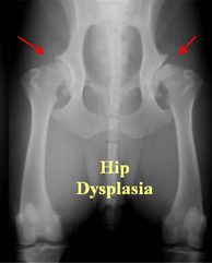
Hip dysplasia is a polygenic and multifactorial disorder influenced by genetics, rapid growth, nutrition, and exercise. It occurs when the femoral head (ball) and acetabulum (socket) fail to develop congruently, leading to joint laxity. This instability causes:
- Subluxation (partial dislocation) of the femoral head
- Abnormal cartilage wear, leading to osteoarthritis
- Remodeling and deformation of the femoral head and acetabulum
Over time, the joint compensates with osteophyte formation (bone spurs) and thickening of the joint capsule, worsening stiffness and discomfort.
Elbow Dysplasia: Pathogenesis and Components
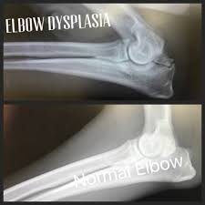
Elbow dysplasia is a complex developmental disorder involving abnormal bone growth, cartilage formation, and joint stress. It comprises several conditions:
- Fragmented Medial Coronoid Process (FCP) – A fracture of the medial coronoid due to uneven pressure distribution in the joint.
- Ununited Anconeal Process (UAP) – Failure of the anconeal process to fuse with the ulna, leading to instability.
- Osteochondritis Dissecans (OCD) – A cartilage defect in the medial humeral condyle, causing joint inflammation.
- Elbow Incongruity – Mismatched growth of the radius, ulna, or humerus, leading to uneven weight distribution.
These abnormalities increase joint stress, leading to cartilage wear, synovial inflammation, and osteoarthritis.
Clinical Signs and Diagnosis
Both conditions present as:
- Lameness, worsening with exercise
- Joint stiffness and pain
- Reluctance to rise, jump, or exercise
Diagnosis involves:
- Radiography (X-rays) – Detects joint incongruities, osteophytes, and subluxation.
- Computed Tomography (CT) or MRI – Provides detailed imaging of bone and cartilage.
- Orthopedic Examination – Assessing joint laxity (e.g., Ortolani test for hip dysplasia).
Treatment and Management
- Surgical Options – Triple Pelvic Osteotomy (TPO), Total Hip Replacement (THR), or Arthroscopic Surgery for elbow dysplasia.
- Medical Management – NSAIDs, weight control, physiotherapy, and joint supplements (glucosamine, chondroitin, omega-3s).
- Breeding Programs – Hip and Elbow Scoring (e.g., BVA, OFA, PennHIP) to reduce genetic prevalence.
Hip and elbow dysplasia require lifelong management, but early detection and appropriate care can significantly improve a dog's mobility and quality of life.
Hip Dysplasia Scoring (BVA/KC Hip Scoring Scheme)
The British Veterinary Association (BVA) Hip and Elbow Dysplasia Scoring System is a radiographic evaluation method used to assess and grade the severity of hip and elbow dysplasia in dogs. It is widely used in the UK and some other countries to guide responsible breeding practices and help reduce the prevalence of these hereditary joint disorders.
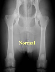
The hip scoring system is based on radiographic evaluation of the hips, typically performed when the dog is at least 12 months old. The BVA panel assesses various anatomical features related to joint conformation and assigns scores based on abnormalities.
Radiographic Procedure
- The dog is sedated or under general anesthesia.
- A ventrodorsal extended-leg hip radiograph is taken.
- The image must be high quality and positioned correctly (pelvis symmetrical, femurs parallel).
Scoring Criteria
The BVA assigns 0–6 points for nine anatomical features of each hip joint, giving a total score of up to 53 per hip and 106 for both hips combined.
- Norberg Angle – Measures femoral head coverage by the acetabulum.
- Subluxation – Degree of looseness in the hip joint.
- Cranial acetabular edge – Shape and conformation of the front edge of the acetabulum.
- Dorsal acetabular edge – Degree of acetabular coverage over the femoral head.
- Cranial effective acetabular rim – Integrity of the acetabular margin.
- Acetabular fossa – Depth and shape of the acetabulum.
- Caudal acetabular edge – Shape and conformation of the rear acetabular margin.
- Femoral head/neck exostosis (remodeling) – Presence of osteophytes (bone spurs) or other abnormalities.
- Femoral head re-contouring – Changes in the shape of the femoral head due to abnormal wear.
Scoring Interpretation
- Total score (both hips combined) ranges from 0 to 106.
- Lower scores indicate better hip conformation (0 = perfect hips, 53+ = severe dysplasia).
- The breed median score is used as a benchmark, and breeding dogs should ideally have scores lower than the breed median.
Elbow Dysplasia Scoring (BVA/KC Elbow Scoring Scheme)
The elbow dysplasia scoring system evaluates degenerative joint changes that indicate elbow dysplasia. It is a 0–3 grading system, with the worst elbow determining the final score.
Radiographic Procedure
- Dogs must be 12 months or older.
- Three high-quality radiographic views are taken:
- **Flexed mediolateral
FCI Hip Dysplasia (HD) and Elbow Dysplasia (ED)Grading System
The Fédération Cynologique Internationale (FCI) Hip and Elbow Dysplasia Scoring System is a radiographic grading method used internationally to assess hip and elbow dysplasia in dogs. Unlike the British Veterinary Association (BVA) scoring system, which assigns numerical values, the FCI system uses a categorical grading scale based on radiographic interpretation.
The FCI hip dysplasia grading system is based on the Norberg angle, joint congruity, acetabular shape, and secondary osteoarthritic changes. It follows a 5-grade classification system from A (normal hips) to E (severe dysplasia).
Radiographic Procedure
- Dogs must be at least 12 months old (large breeds may require evaluation at 18 months).
- Ventrodorsal extended-leg hip radiographs are taken with:
- Pelvis symmetrically positioned
- Femurs parallel
- Patellae centered over the femoral shafts
- The Norberg angle is measured (≥105° is ideal).
Grading Criteria
Breeding Recommendations
- Dogs with Grade A or B are considered suitable for breeding.
- Dogs with Grade C may be bred with an A-graded dog but should be avoided in high-risk breeds.
- Dogs with D or E grades should not be bred.
The FCI elbow dysplasia scoring system assesses joint congruity, osteoarthritis (OA), and primary lesions such as:
- Ununited Anconeal Process (UAP)
- Fragmented Medial Coronoid Process (FCP)
- Osteochondritis Dissecans (OCD)
- Joint incongruity
Radiographic Procedure
- Dogs must be at least 12 months old.
- Mediolateral elbow radiographs are taken in a flexed and extended position.
- The presence of osteophytes (bone spurs) is the main criterion.
Grading Criteria
Breeding Recommendations
- Only Grade 0 elbows should be bred.
- Grade 1 may be acceptable in some cases.
- Dogs with Grade 2 or 3 should not be bred.
Key Differences Between FCI and BVA Scoring Systems
The FCI system is widely used in Europe, South America, and Asia, while the BVA system is standard in the UK and Commonwealth countries.